Scientific Context
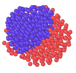
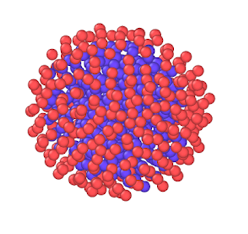
Figure 1: MD snapshot of an AgCo Janus and core-shell nanoalloy
The rich versatility of nanoalloys makes them suitable for a large range of prospective applications, with examples in catalysis, plasmonics, sensing, and data storage. The advantages of nanoalloys are linked to their small size that achieves high surface area available for catalysis with only small amounts of often costly materials, such as Rh, Pd, or Au. Even more so, the number of atoms at surface steps, which may represent important reactive sites, is greatly enhanced in nanoparticles (NPs). In nanoalloys, the amount of rare raw material may be further decreased by replacing the core of the particles with another material. Furthermore, the second material may be used to tune the electronic structure at the particle surface. Even more possibilities arise with multi-component particles.
The realization of the full potential of these systems requires the design of nanoalloys of controlled size, morphology, and chemical composition. Over the last decades, a considerable number of physical and chemical synthesis methods have been developed, allowing now for some control of the final product. In this context, the path dependence, i.e. the synthesis pathway, and the environment, such as the contact with a substrate, is crucial because of the inherent metastability of nanoalloys. The wealth of structural possibilities of nanoalloys calls for reliable methods for their characterization.
Alongside X-ray scattering methods, electron microscopy stands out as a powerful analysis tool. High-resolution transmission electron microscopy (HRTEM) allows for imaging at atomic resolution in case of aberration corrected microscopes. Scanning transmission electron microscopy (STEM) in the high-angle annular dark-field mode (HAADF), in particular, is useful for imaging alloyed systems because of the contrast regarding atom types. If one wants to fully analyze the distribution of sizes, shapes, and chemical orderings of a given sample, many images need to be recorded and analyzed for a representative statistical analysis. Selecting only a small portion of the sample for further analysis can also introduce some subjectivity into the final results.
Project Approach
Recent developments in electron microscopy allow the observation of nanomaterials at atomic resolution. The interpretation of images from experiments is, however, not always straightforward because of lens aberrations. Usually, image simulations are used to compare with images from experiments and to determine structural details. This process is very time-consuming for electron microscopy specialists. With this project, we want to make the analysis of large sets of electron microscopy images practical and maybe even more objective, thanks to automated systems based on deep learning (DL).
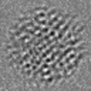

Figure 2: Simulated HRTEM image of an AgCo Janus and core-shell nanoalloy
To establish a training database for the envisioned DL systems, we will rely on NP structures obtained from atomistic simulations. As we concentrate here on nano-alloys, interatomic potentials for transition metals are required as a basis for the simulations. The second moment approximation of tight-binding (TB-SMA) has been used successfully in the past for many studies on bi-metallic nano-alloys. With these efficient interatomic potentials, it is relatively easy to obtain a sufficient number of realistic structures of nanoalloy particles by sampling the correct thermodynamic ensemble. The high degree of control during the simulations makes it possible to sample particles in meta-stable configurations, e.g. core-shell systems, that are partially ordered or in Janus configuration as obtained experimentally in certain growth conditions. These structures can then be used as a basis for the generation of electron microscopy images. While these kinds of calculations, based on the multi-slice technique, have been used for a long time to generate images for comparison with images from experiments, recent software improvements make the automatic and fast generation of many realistic images more practical.
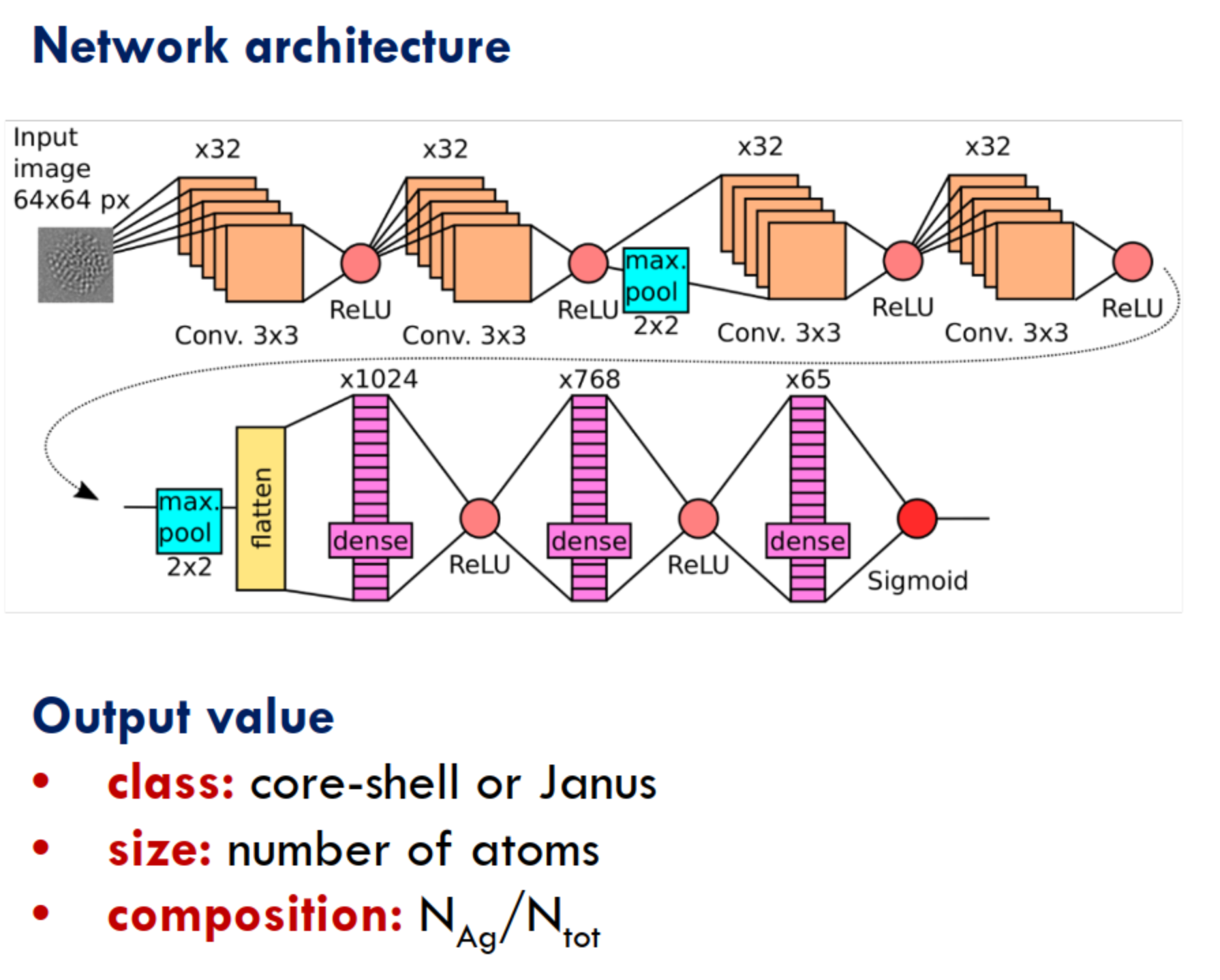
Figure 3: Schematic of a convolutional neural network applied to nanoalloy HRTEM images
DL is a powerful tool for image analysis, and is becoming rapidly a main-stream technique in numerical physics, too. Our project obviously benefits from these developments in several respects: Firstly, from a fundamental perspective, many types of DL have been established for various tasks that are relevant for this project, such as image analysis, image classification, and image enhancement. Secondly, much empirical evidence guiding the selection and parametrization of particular DL architectures has been accumulated recently within the machine learning community.
Objectives
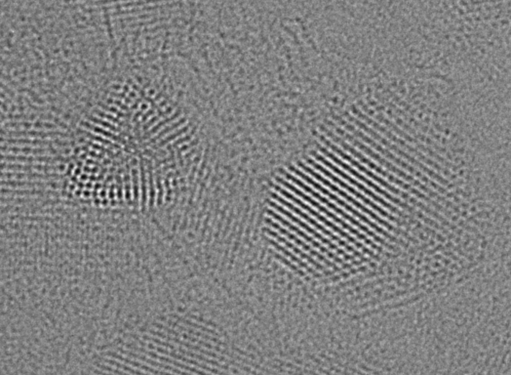
Figure 4: Experimental HRTEM image of a AgCo nanoalloy
With this project, we wish to seize the emerging opportunity of leveraging DL for the analysis of atomic resolution images of nano-objects that arises thanks to recent developments in three relevant fields: Firstly, electron microscopes (STEM and HRTEM) has reached atomic resolution. Secondly, atomistic simulations of nano-objects are now realistic and fast enough to be able to generate databases for the subsequent training of DL systems. Thirdly, while DL remains a very active field of fundamental research, it has become efficient and versatile enough to be used in practical applications.
These DL systems will lead to:
- Higher quality images (denoising, super-resolution)
- Automated analysis of nanoalloy properties (number, size, shape, crystallinity, (meta-)stability, etc.)
- Classification of nanoalloys in terms of chemical ordering
The project coordinator has already proven the feasibility and potential of this approach in a recent publication on the classification of HRTEM images of carbon nanotubes. A web interface for this analysis is available at https://hrtem-analysis.fr.